Anita Markbåge medverkar i Länsradion i Örebro och pratar om elöverkänslighet. Anita delar personliga berättelser och information om tillståndet och diskuterar även filmen Electric Malady, regisserad av Marie Lidén, som ...
Under en tioårsperiod reste konstnären och filmskaparen Marie Lidén regelbundet till Närke för att skildra Williams liv. Filmen bjuder in publiken till Williams isolerade värld, en ung man som motvilligt ...
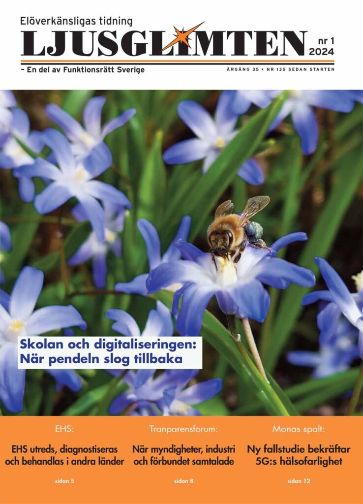
Nya numret av Ljusglimten har publicerats. Det innehåller en blandning av aktuella nyheter, värdefulla råd, intervjuer, mat och annat i stort och smått. Bli medlem så får du fyra nummer ...
Det sprids i våra medier och på sociala medier myter om att EHS/elöverkänslighet är en psykisk sjukdom som kan botas genom kognitiv beteendeterapi (KBT). Att patienten ska träna sig att ...
Det sprids i våra medier och på sociala medier myter om att provokationstester inte fungerar för att diagnostisera EHS. Provokationstesterna är i många fall en viktig del i de tester ...
Bevisen på samband är tydliga i forskning och verkliga livet. För att belysa de tydliga kopplingar som finns mellan EHS/elöverkänslighet och påverkan från elektromagnetiska fält (EMF), t.ex. el- och magnetfält ...
Denna webbplats använder kakor för bästa möjliga användarupplevelse. Godkänn innan du fortsätter. Jag godkännerLäs mer...
Integritetspolicy
Privacy Overview
This website uses cookies to improve your experience while you navigate through the website. Out of these, the cookies that are categorized as necessary are stored on your browser as they are essential for the working of basic functionalities of the website. We also use third-party cookies that help us analyze and understand how you use this website. These cookies will be stored in your browser only with your consent. You also have the option to opt-out of these cookies. But opting out of some of these cookies may affect your browsing experience.
Necessary cookies are absolutely essential for the website to function properly. This category only includes cookies that ensures basic functionalities and security features of the website. These cookies do not store any personal information.
Functional cookies help to perform certain functionalities like sharing the content of the website on social media platforms, collect feedbacks, and other third-party features.
Performance cookies are used to understand and analyze the key performance indexes of the website which helps in delivering a better user experience for the visitors.
Analytical cookies are used to understand how visitors interact with the website. These cookies help provide information on metrics the number of visitors, bounce rate, traffic source, etc.
Cookie
Varaktighet
Beskrivning
_ga
1 year 1 month 4 days
Google Analytics sets this cookie to calculate visitor, session and campaign data and track site usage for the site's analytics report. The cookie stores information anonymously and assigns a randomly generated number to recognise unique visitors.
_ga_*
1 year 1 month 4 days
Google Analytics sets this cookie to store and count page views.
_gcl_au
3 months
Google Tag Manager sets the cookie to experiment advertisement efficiency of websites using their services.
sbjs_current
session
Sourcebuster sets this cookie to identify the source of a visit and stores user action information in cookies. This analytical and behavioural cookie is used to enhance the visitor experience on the website.
sbjs_current_add
session
Sourcebuster sets this cookie to identify the source of a visit and stores user action information in cookies. This analytical and behavioural cookie is used to enhance the visitor experience on the website.
sbjs_first
session
Sourcebuster sets this cookie to identify the source of a visit and stores user action information in cookies. This analytical and behavioural cookie is used to enhance the visitor experience on the website.
sbjs_first_add
session
Sourcebuster sets this cookie to identify the source of a visit and stores user action information in cookies. This analytical and behavioural cookie is used to enhance the visitor experience on the website.
sbjs_migrations
session
Sourcebuster sets this cookie to identify the source of a visit and stores user action information in cookies. This analytical and behavioural cookie is used to enhance the visitor experience on the website.
sbjs_session
1 hour
Sourcebuster sets this cookie to identify the source of a visit and stores user action information in cookies. This analytical and behavioural cookie is used to enhance the visitor experience on the website.
sbjs_udata
session
Sourcebuster sets this cookie to identify the source of a visit and stores user action information in cookies. This analytical and behavioural cookie is used to enhance the visitor experience on the website.
Advertisement cookies are used to provide visitors with relevant ads and marketing campaigns. These cookies track visitors across websites and collect information to provide customized ads.
Cookie
Varaktighet
Beskrivning
test_cookie
15 minutes
doubleclick.net sets this cookie to determine if the user's browser supports cookies.